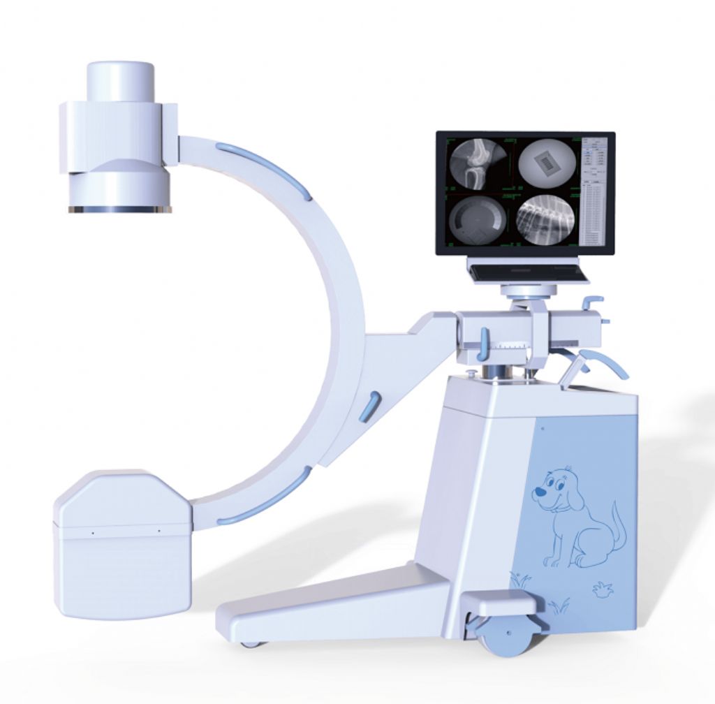
Description
Configuration
1- New (With electric auxiliary support arm) C-arm Frame
2- High-frequency high-voltage x-ray generator and high-frequency inverter power supply
3- 24 inch ordinary LCD, resolution 1920×1200
4- 9 inch three field image intensifier
5- Megapixel ultra-low illumination digital camera, Camera pixel matrix description: 1024×1024
6- Digital acquisition and processing system
7- Dense grid 40L/cm grid ratio: 8:1 focal length: 90C
8- Electric adjustable beam collimator
9- Handheld controller
10- Laser cross positioner
Features
1. High quality combined high-frequency and high-voltage X-ray generator, greatly reducing the X-ray exposure;
2. It has the function of automatic tracking of perspective kV and Ma, which makes the image brightness and clarity in the best state automatically;
3. The host operation interface of the human graphical LCD touch screen is adopted to make the operation more intelligent and convenient;
4. The design of the hand-held controller makes the operation of the instrument more convenient;
5. The 9-inch three field image intensifier is used, with stable and reliable quality and good image definition; 6. The megapixel ultra-low illumination digital camera is used, with clearer image;
7. Standard workstation and advanced image software processing technology make the image clearer, convenient for doctors’ operation and diagnosis, standard DICOM interface and easy to link with hospital information system;
8. New frame design, small and beautiful appearance;
9. Realize the function of digital photographing, make the photographing operation more convenient and the image digital processing more efficient
Product Specification
1- Monoblock
1.1 Focus:0.6/1.8
1.2 Anode capacity:35.5kJ(47kHu)
1.3 Tube Heat capacity:650kJ(867kHu)
1.4 Power output:5kW
1.5 Inverter Frequency:≥40kHz
1.6 Continous Fluoroscopy (Manual/automatic)
1.6.1 Tube voltage:40kV~120kV
1.6.2 Tube current:0.3mA~4mA
1.6.3 Automatic brightness tracking function
1.7 Pulse fluoroscopy
1.7.1 Tube voltage:40kV~120kV
1.7.2 Tube current:0.3mA~30mA
1.8 Radiography mode
1.8.1 Radiography:40kV~120kV
1.8.2 Radiography tube current:25mA~100mA
1.8.3 Radiography mAs:1.0mAs~180mAs
1.9 Beam limiter: Electric iris + linear symmetrical rotatable
1.10 Working environment conditions
1.10.1 Environment temperature :10°C—40°C
1.10.2 Relative humidity:30%—75%
1.10.3 Atmospheric pressure:700hpa—1060hpa
1.11 Operating power condition
1.11.1 Power supply voltage and phase number: single-phase 220V ± 22V
1.11.2 Power frequency:50Hz±1Hz
1.11.3 Internal resistance of power supply: no more than 1 Ω
2- Imaging System
2.1 Image intensifier: 9 ″ three field e5764sd-p3, center resolution 4.8lp/mm
2.2 Ultra low illuminance, megapixel black and white progressive scanning camera pixel matrix description: 1024×1024
2.3 LCD: 24 inch ordinary LCD, resolution 1920×1200, working frequency: 60Hz
2.4 Image acquisition and processing workstation
2.4.1 Registration: Registration preservation, medical record query, worklist
2.4.2 Acquisition: start acquisition, prepare recording, reset, horizontal mirror, vertical mirror, window adjustment, magnifying glass, negative image, edge enhancement, recursive noise reduction
2.4.3 Processing: four windows, nine windows, sharpening, horizontal mirror, vertical mirror, text annotation, length measurement
2.4.4 Report: save, preview, expert template
2.4.5 DICOM function: DICOM browsing, network service
2.5 Image definition index
2.5.1 Gray level: ≥ 11
2.5.2 Line pair resolution: ≥ 2.0LP/mm
3- Mechanical Part
3.1 Forward and backward movement: 200mm
3.2 Rotation around horizontal axis: ± 180 °
3.3 Rotation around vertical axis: ± 15 °
3.4 Focal screen distance: 960mm
3.5 C-arm opening: 760mm
3.6 Arc depth of C arm: 640mm
3.7 Sliding along the track: 120 ° (+ 90 ° ~ – 30 °)
3.8 Electric lifting of column: 400mm
3.9 Guide wheel and main wheel: the guide wheel can rotate in any direction, and the main wheel can rotate ± 90 °
3.10 Monitor on frame rotation ≥ 300 °
3.11 Light thrust


Reviews
There are no reviews yet.