Description
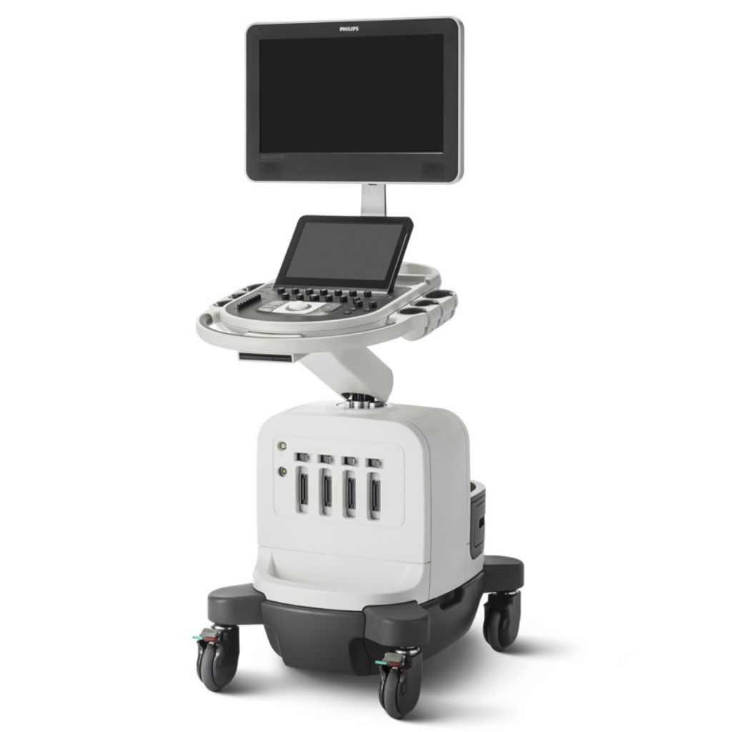
Choosing a new ultrasound system is all about balance. You need accurate diagnostic information quickly, a simplified yet intuitive user interface, and easy access to critical features, along with an ergonomic design and the latest technology.
Philips Affiniti 50 Features
The following are the standard features of an Affiniti 50.
21.5” LCD display with four-way articulation
12” widescreen tablet-like touchscreen
4 transducer ports and one-handed transducer access
Intelligent software architecture
Combined 512 GB storage capacity
DVD+RW drive
Wireless networking
State-of-the-art ergonomic designed system cart for comfort and convenience
Easy-to-learn graphical user interface with reduced number of hard controls
Advanced control panel design with fewer, clustered controls
2D mode
Tissue Harmonic Imaging(ThI)
M mode
Color Doppler
Color Power Angio Doppler
Pulse wave Doppler
Auto color and auto Doppler
Transport mode : The system lasts 45 minutes before recharge
PureWave crystal technology
Auto Doppler for vascular imaging
SonoCT real-time compound imaging
XRES adaptive image processing
iSCAN intelligent optimization
AutoSCAN intelligent optimization
iOPTIMIZE intelligent optimization
Tissue aberration correction (TAC)
Coded beamforming
QuickSAVE feature
Cineloop review: up to 2,200 frames of 2D and color images
High Q automatic Doppler analysis
Philips Affiniti 50 Technology Definitions
SonoCT: Real-time Compound Imaging on the Philips Affiniti 50 that obtains multiple coplanar, tomographic images from different viewing angles, then combines them into a single compound image at real-time frame rates.
XRES adaptive image processing: Real-time speckle reduction standard on the Affiniti 50 that also enhances edge definition.
AutoSCAN: This Philips Affiniti 50 feature automatically and continuously optimizes the brightness of the image at the default gain and TGC settings for the best image display. It can be turned on and off.
iSCAN intelligent optimization: Automatic one-button global image optimization standard on the Philips Affiniti 50 through AI adjustment of TGC, Doppler and receiver gain, compression curve, Doppler PRF, and Doppler baseline.
QLAB quantification software: QLAB is an onboard quantification program that can also be used on a computer to analyze data. QLAB on the Philips Affiniti 50 has the following available plugins: 3DQ, IMT, FHN, MVI, EA, VPQ and ROI.
QLAB 3DQ GI: This QLAB tool on the Philips Affiniti 50 allows viewing, quantification, cropping, rotation, and measurements of 3D image data sets.
QLAB IMT: This Affiniti 50 QLAB tool makes measurement of the intima media thickness in carotids and superficial vessels quick and consistent.
QLAB MVI: MicroVascular Imaging on the Philips Affiniti 50 maps contrast agent progression, measuring frame-to-frame changes, suppressing background tissue and capturing additional data that make it significantly easier to visualize the vessels.
QLAB VPQ: Vascular Plaque quantification is an application on the Philips Affiniti 50 that uses 3D technology to examine arteries and determine the risk of stroke or cardiovascular disease, measure how much plaque is present and the percentage of vessel reduction.
QLAB ROI: A plugin within QLAB on the Affiniti 50 that uses contrast and 2D imaging to increase the consistency and reliability of acoustic measurements.
a2DQA.I.: Automated Cardiac 2DQ Quantification is an available aEF/RAC workflow and aTMAD (Automated Tissue Motion Annular Displacement) workflow. As an aEF/FAC workflow it uses the latest generation of 2D speckle tracking technology to compute the left ventricle global volume and area analysis from 2D single images. The volume measurements are made based on Simpson’s Single Plane Method of Disks (MDO). The aTMAD workflow provides value annular displacement curves over time and tracks mitral valve and annular motion in other valves over time.
aCMQA.I.: Automated Cardiac Motion 2D Quantification on the Philips Affiniti 50 is an available global strain workflow providing; volume EF and area FAC, Longitudinal strain and strain rate, circumferential strain and strain rate, radial and transversal displacement, radial fractional shortening, global rotation, and more. A user-definable workflow is also included. The aCMO supports the functions of a2DQ.
Philips Affiniti 50 ports
4 active transducer ports
DICOM
Ethernet port
HDMI port
USB port
2 on-board peripheral connections via USB
Philips Affiniti 50 Imaging Modes
B-mode
M-mode
(PW) Spectral Doppler
Auto color and auto Doppler
Steerable continuous wave (CW) Doppler
Tissue Doppler Imaging (TDI/TDI PW)
3D/4D and MPR imaging (hybrid transducers)
Freehand 3D volume and MPR imaging
Spatio-Temporal Image Correlation (STIC) imaging
Panoramic imaging
Contrast imaging – cardiovascular
Contrast imaging – general imaging
Interventional imaging
2D imaging
Tissue Harmonic Imaging (THI)
Color Doppler
Color Power Angio imaging (CPA)
Strain-based elastography
4D imaging
Duplex mode
Triplex mode
Dual imaging
Philips Affiniti 50 Applications
Applications or Apps are the types of exams or studies that an ultrasound machine can do. More than this if an ultrasound machine supports a specific application it will have calculations, measurement and reporting software included to support those apps and make them useful in a clinical environment.
The Philips Affiniti 50 is a true shared-service ultrasound machine and offers the broadest selection of applications possible.
Abdominal
Obstetrical
Fetal echo
Cerebrovascular
Vascular (peripheral, cerebrovascular, temporal TCD, and abdominal)
Abdominal vascular
Gynecological and fertility
Small parts and superficial
Musculoskeletal
Pediatric general imaging
Prostate
Echocardiography (adult, pediatric, fetal)
Stress echocardiography
Transesophageal echocardiography (adult and pediatric)
Surgical imaging
Interventional imaging
Contrast imaging
Bowel imaging
Strain elastography
Shear wave elastography
Perioperative
Epicardial echocardiography


Reviews
There are no reviews yet.