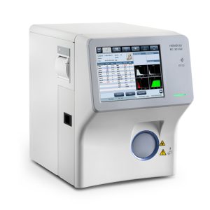
Mindray BC-60 R Vet Advanced Veterinary Hematology Analyser
72.580,00 د.إ
Description
With the deepening of the concept of animal health, people pay more attention to their pet’s health, and veterinarians have higher requirements for diagnostic equipment. The veterinary hematology analyzer, as one of the core equipment in the laboratory department, is assuming greater responsibility.
Exponentialgrowth of pathological samples in animal hospitals requires accurate test results;
Veterinary disease diagnosis requires more comprehensive clinical parameters, such as reticulocyte series parameters for the evaluation of animal anemia types;
Veterinarians and laboratory personnel need intelligent equipment, intuitive results, and convenient operation;
According to the characteristics of veterinary hematology analysis, Mindray Animal Medical insists on taking technological innovation as the basis, taking customer needs as the goal, and taking the clinical characteristics of animals as guidance. Now, we have developed hematology analyzer products that are suitable for animals, and we have become a pioneer in the field of a new generation of hematology analyzers.
SF-Cube Veterinary Hematology Analysis Platform
3D Cube Analysis Technology Combining Scattered Light and Fluorescence Dyeing
S-Scatter: forward laser scatter and side laser scatter to detect the size and complexity of cells F-Fluorescence: side laser scatter fluorescence detection of nucleic acid content in cells
Cube-3D cubic analysis technique combining both scattered light and fluorescence.
The analysis technology platform dedicates to veterinary needs. BC-60R Vet is based on 3D analysis technology, improves the accuracy of leukocyte classification, and can find more abnormal cells (i.e., Band, nucleated red blood cells), providing more reference value for clinical practice.
3D Scattergram of WBC 3D Scattergram of RET
The Effect of the 3rd Generation Fluorescent Dyeing Technology is Better
The Development of Fluorescent Dyeing Technology
2010 2017 2022
About 2010, the dyeing technology based on the human medical platform was applied in veterinary blood cell dyeing
2nd generation fluorescent dyeing technology: about 2017, the signal-to-noise ratio of leukocyte
DNA dyeing and the signal-to-noise ratio of RNA in reticulocyte testing;
3rd generation fluorescent dyeing technology: combining 2nd generation technology and SF-Cube platform, Mindray Animal Medical developed the animal-specific technology for veterinary leukocyte differentiates and reticulocyte recognition.
Comparison of Different Dyeing Technology
represents better performance
| Fluorescent Dyeing Technology 1.0* | Fluorescent Dyeing Technology 2.0* | Fluorescent Dyeing Technology 3.0* | |
| The Signal-to-Noise Ratio of Leukocyte DNA |
|
|
|
| Differentiation of Lymphocyte and Neutrophil in Abnormal Animal Samples |
|
|
|
| The Signal-to-Noise Ratio of Reticulocyte RNA |
|
|
Dyeing technology combined with SF cube platform technology is the core technology of hematology analysis. With the continuous improvement of animal clinical requirements for test results, the traditional fluorescent dyeing technology has inaccurate results in the accuracy of leukocyte differentiation and a reticulocyte count of abnormal samples. The 3rd generation fluorescent dyeing technology overcomes traditional technology’s barriers and improves the accuracy of leukocyte differentiation and reticulocyte count.
Better Differentiation of Lymphocyte and Neutrophil
The traditional dyeing technology has the problem that it is challenging to distinguish Lymphocyte cells from Neutrophil, resulting in the abnormal differentiation of leukocytes. The 3rd generation of dyeing technology is better at differentiating Lymphocyte and Neutrophil and more accurate to recognize and classify the leukocytes.
Clear Differentiation between Lymphocyte and Neutrophil
Reticulocyte More Accurate
In distinguishing the type anemia and the degree of anemia, reticulation is a very important parameter. The 1st generation dyeing technology has limitations in specificity and anti-interference ability, which affects the accuracy of test results of abnormal samples and is not conducive to the clinical diagnosis and treatment of animals.
WBC RET PLT
High sensitivity optical detection system and a new high-specific fluorescent dye could significantly improve the performance of RET channel detection.
Technical Specifications
Principles
Flow Cytometry and SF Cube* method to count WBC 5-part, RET Sheath Fluid Impedance method for counting RBC and PLT, Optical-PLT counting
Cyanide free reagent for hemoglobin test
*S: Scatter; F: Fluorescence; Cube: 3D analysis
Animal species
Phase I: cat, dog and horse;
Phase II: rat, mouse, rabbit, monkey and pig;
Phase III: cow, ferret, goat, sheep, camel, llama, panda, alpaca); 16 Animal species+50 self-define animal species;
Parameters(33 Parameters)
WBC Series: WBC, Neu(#,%), Mon(#,%), Lym(#,%), Eos(#,%), Bas(#,%) (11 Items) RBC Series: RBC, HGB, HCT, MCV, MCH, MCHC, RDW-SD, RDW-CV (8 Items)
RET Series: RET(#,%), IRF, LFR, MFR, HFR, RHE (7 Items) PLT Series: PLT, PDW, MPV, P-LCR, P-LCC, PCT, IPF (7 Items)
Sample Type
Whole blood、Predilute*(rat, mouse)
Sample Volume
Whole blood (CBC+DIFF+RET) 34uL Predilute*: 20uL
Throughput
Up to 40 samples per hour (CBC+DIFF+RET)
Operating Environment
Temperature: 10°C~30°C Humidity: 30%~85%
Air pressure: 70 kPa~106 kPa Voltage: 110V-220V
Display & Interface
12 inch TFT Touch Screen USB, LAN
Support bi-directional LIS
Dimension and Weight
Analyzer
Platelet Detection System
Two methods: PLT-I、PLT-O

